 International Patients
Biopsy
Targeted biopsy for prostate
Targeted biopsy for prostate combines magnetic resonance imaging (MRI) with real-time ultrasound images so that physicians can precisely track, target, and sample the prostate tissue that appears to be suspicious. Using a fusion biopsy may mean that you can undergo fewer biopsies. These biopsies can be performed both With anesthesia and with local anesthesia.
How It Works
We perform a targeted biopsy using the state-of-the-art UroNav® system to diagnose prostate cancer. This technique fuses highly detailed magnetic resonance imaging (MRI) with real-time ultrasound using the Artemis device. The procedure typically takes 15 to 20 minutes in our office under local anesthesia. It provides highly accurate information about the location of the cancer and its relation to nerves and sphincters.
Targeted Biopsy with UroNav® system

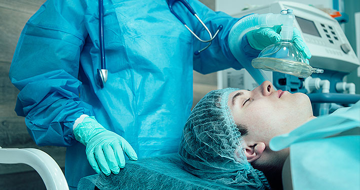
“Twilight Anesthesia” (Conscious Sedation)
Conscious sedation is a service offered to our patients for their comfort. Both systematic and MRI fusion guided prostate biopsies can be performed in the office at 625 Madison Avenue with the assistance of an anesthesiologist who is able to give intravenous medications to gently and mildly sedate patients so that they can better tolerate any potential discomfort.
This sedation can be mild or moderate based on the needs of the patient or the dosage determined appropriate by the anesthesiologist. Patients can respond to commands but will likely fall asleep through most of the procedure. Since patients are not unconscious, there is no need for external breathing support.

While patients can choose if they would like IV sedation, all patients receive local anesthesia (usually injectable lidocaine) at the site of the biopsy. It takes approximately 30 minutes for sedation to wear off.
A team member monitors the patient after the procedure to make sure he is walking comfortably and will be discharged from our office in stable condition. We ensure that all patients urinate on their own at least once prior to leaving our clinic post-biopsy.
Targeted biopsy for prostate
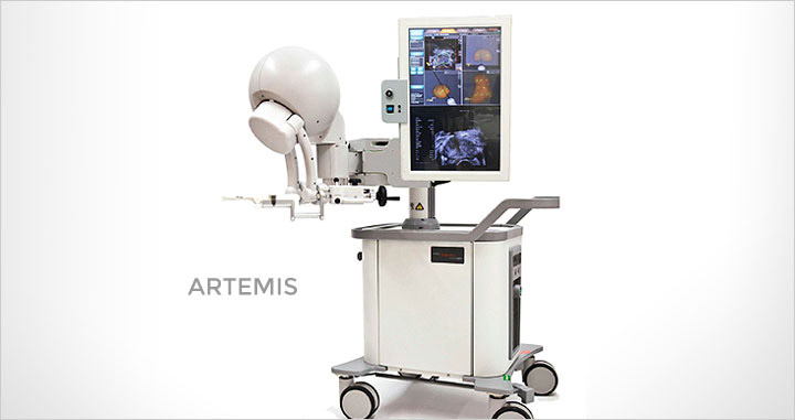
Artemis
Artemis is an imaging device that allows 3D prostate visualization and tracking. The Artemis biopsy diagnostic procedure combines traditional biopsy with advanced imaging to determine the presence of prostate cancer.
This technique is done in two steps.
First an MRI is done, and the scans are loaded onto software allowing the radiologist to mark the prostate gland and the regions of interest for biopsy. This is known as segmentation. This information is then loaded onto Artemis for a targeted biopsy. The procedure can be done in an outpatient setting under local anesthesia and takes only a few minutes.
MRI-targeted biopsies via the transperineal approach
We perform 400-450 MRI-targeted biopsies every year through the rectum.
In the future, we expect to conduct MRI-targeted biopsies via a Transperineal approach (a thin needle is inserted through the skin of the perineum and into the prostate). There are currently no studies directly comparing the different MRI-targeted approaches, however, there is evidence that the transperineal approach has excellent detection rates with none of the septic complications that can occur after the rectal approach.
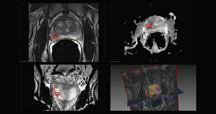
MRI guided fusion biopsy planning targets